Promising nanoscale material captured by electron microscopy

When you go on a nanoscale expedition, it’s important to get good photos. Scientists creating new chemicals and materials at the level of nanometers — 1/100,000 the width of a human hair — require high-resolution images of their atomic structure to confirm new synthesis methods and explore potential applications.
For a recent study published in Science, University of Illinois Chicago researchers visually captured the structure of MXenes, synthetic compounds with promise for energy storage, sensing and superconductivity. The collaboration with scientists from the University of Chicago drew upon the world-class electron microscopy resources and expertise at UIC to reveal novel structures with intriguing properties.
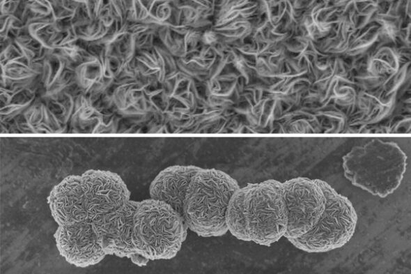
Images collected by Robert Klie, UIC professor and head of physics, and physics graduate student Francisco Lagunas captured MXenes in layered “carpets” of atoms as well as in a previously unseen ruffled sphere shape. Those “fluff balls,” as Klie describes them, were notable enough to make the cover of the March 24 issue of Science.
“With these new properties, we can really control the quality as well as functionality of those materials,” Klie said. “This new paper comes up with a way to not only have these clean, well-controlled materials, but also grow them in a way such that it’s now possible to manufacture in large quantities.”
Since the discovery of MXenes in 2012, materials scientists and chemists have experimented with how best to create these unique compounds to maximize their utility. The original method involves a slow, harsh etching process that generates hazardous waste, creating an obstacle for scaling these materials from the laboratory to industrial production.
In the new Science paper, researchers in the laboratory of Dmitri Talapin at University of Chicago developed two faster and cleaner processes of making MXenes using extreme heat (up to 950 degrees Celsius) or chemical vapor. Researchers then needed to visually confirm that the MXenes produced by these new methods carried the desired properties and whether they displayed novel characteristics.
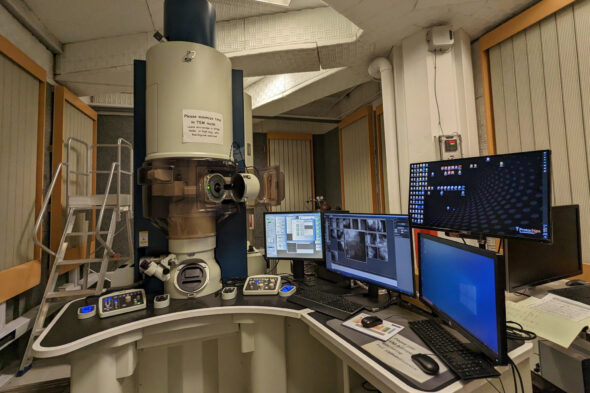
To do so, they turned to UIC and the Electron Microscopy Core, with its state-of-the-art scanning transmission electron microscopes. Klie and Talapin’s groups have a long-standing collaboration, including a preceding 2020 Science paper on creating MXenes with customized functional groups that can change their abilities.
“We really need to understand what atoms are where, and how this correlates to the properties,” Klie said. To that end, Lagunas developed new microscopy approaches for studying the growth and structure of delicate compounds 100,000 times thinner than a sheet of paper.
“Francisco worked on a lot of novel methods to provide information on the atomic structure and where the functional groups sit on the surface and how they arrange,” Klie said.
One innovation was the ability to slice through the MXene spheres in order to view them in cross-section, allowing scientists to examine the core of these complex structures and infer how they form. While these experiments were performed at the Canadian Centre for Electron Microscopy, the UIC core has since added this type of focused ion beam electron microscope.
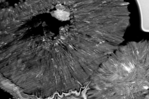
“We used a focused ion beam to cut out a small section of the MXene sphere, and you can see that the inside appears to be hollow, with small titanium carbide crystals that form in the center,” Lagunas said. “We think that those crystallites might act as some sort of nucleation point for the growth of the MXenes.”
As the MXene collaboration continues, the exceptional imaging abilities of the UIC team expands. In 2024, they will add the world’s first electron microscope with a magnetic field-free objective lens, via a $4 million award from the National Science Foundation’s Major Research Instrumentation Program.
Talapin’s group will be one of many from universities around the world expected to collaborate with UIC in using the new instrument to advance science. It will also support Klie’s own research on novel materials for solar cells, batteries and other applications in his Nanoscale Physics Group.
“Many materials like superconductors or magnetic materials, when they sit in a very high magnetic field, the properties change,” Klie said. “We will be able to image these materials in a field-free environment with atomic resolution, and then we can apply a small magnetic field and see how it affects a material’s properties. It will improve our ability to collect chemical information by at least a factor of ten.”
