New Alzheimer’s test may identify those at risk

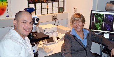
Mary Jo LaDu, associate professor of anatomy and cell biology, and postdoctoral research associate Leon Tai. Photo: Reginald Smith
A pair of new assays that can detect key Alzheimer’s molecules in human brain tissues and in lab mice may offer a quick way to identify new effective drug treatments.
The assays may also prove useful in identifying people at increased risk for the disease, which would help determine who should get a preventive drug should one be discovered, says Mary Jo LaDu, associate professor of anatomy and cell biology at the University of Illinois at Chicago College of Medicine, who is principal investigator of the study.
The study is “paper of the week” in the Feb. 22 issue of the Journal of Biological Chemistry.
A team led by LaDu developed a pair of assays able to detect “mechanistic biomarkers” of Alzheimer’s — a term they coined, to indicate that the tell-tale biomolecules are directly involved in the disease process.
It may also be possible to use the mechanistic biomarkers to track progression of the disease, LaDu says, which is key to studying a disease like Alzheimer’s that takes years or decades to produce symptoms. With no easy way to determine whether progression has been slowed or stopped, researchers have so far failed to find an effective therapy — and failures in clinical trials are expensive.
Two months ago, LaDu’s group reported the development of a new mouse model of Alzheimer’s. The mice carry a human gene that — in its naturally occurring variations — is a major determinant of risk. The new assays, LaDu says, can be used in that animal model to rapidly and cheaply screen large numbers of chemicals for therapeutic effect before any are tested in patients.
One of the hallmarks of Alzheimer’s is the appearance in brain tissue of dense plaques formed by clumps of a protein called amyloid beta. But recent evidence, LaDu says, indicates that smaller, soluble “oligomers” of amyloid beta, rather than the plaques, are responsible for nerve cell death in the disease.
A protein in cells called apoE was long suspected to be involved. One naturally-occurring variant of apoE, called apoE4, is the most important known genetic risk factor for Alzheimer’s disease. People carrying the E4 gene face a 15-times higher risk for Alzheimer’s than the population at large.
Leon Tai, UIC research assistant professor in anatomy and cell biology and first author of the new study, says the team wondered: might healthy apoE be responsible for clearing out toxic amyloid beta oligomers from the nerve cell?
The team developed a pair of assays, each able to distinguish oligomers from plaques. One assay measures the total amount of oligomers — whether bound to apoE or free in solution. The other measures only oligomers bound to apoE.
Using their mice carrying a human apoE gene, the researchers made an important discovery: In mice carrying a robust version of apoE, most oligomers were bound to apoE. But in mice with the E4 gene, most oligomers remained on the loose — apparently the apoE4 protein doesn’t hold on to them very well.
“This is exactly what we expect to see if our hypothesis is correct,” said LaDu. “We think that one of the things that apoE is doing is clearing the soluble neurotoxic oligomers. So this was an exciting result.”
The researchers then turned to brain tissue and cerebrospinal fluid from humans, comparing Alzheimer’s brains to normal controls. Alzheimer’s brains had more free oligomers, while the normal brains had more oligomers bound to apoE — consistent with the idea that the oligomers may be toxic to nerve cells if apoE does not clear the oligomers away.
The team also compared tissue from individuals carrying genes for the different versions of apoE. Again, they found that in the brains of people who had the high-risk E4 gene, there were more oligomers free in solution than in those who had any of the lower-risk versions of apoE.
All results were consistent with their hypothesis that apoE is involved in clearing neurotoxic oligomers of amyloid beta from the brain.
“We are now testing the (assay’s) predictive value for the progression of Alzheimer’s — distinguishing mild cognitive impairment from early and late stage disease,” LaDu said. The ultimate goal, she said, is to identify those at risk before the disease process has begun to cause cognitive decline.
“We will also be looking at using our mouse model for pre-clinical drug development,” LaDu said, using the assays to rapidly screen for compounds that increase the clearing of amyloid beta oligomers from the mice.
Co-authors are Lisa Jungbauer, Stephen Roeske, Katherine Youmans, and Chungjiang Yu of UIC; Tina Bilousova and Karen Hoppens Gylys of the UCLA School of Nursing; Mary S. Easton of Alzheimer’s Research, Los Angeles; Wayne Poon and Lindsey Cornwall of the University of California, Irvine; Carol Miller of the University of Southern California; Harry Vinters of the UCLA School of Medicine; Linda Van Eldik, David Fardo and Steve Estus of the University of Kentucky; and Guojun Bu of the Mayo Clinic in Jacksonville, Fla.
The research was supported by National Institutes of Health grants P01AG030128, AG27465, and AG18879; and grants from the Alzheimer’s Association, the UIC Center for Clinical and Translational Science, the Alzheimer’s Drug Discovery Foundation, and the Daljit S. and Elaine Sardaria Chair in Diagnostic Medicine.
