How do neural support cells affect nerve function?
Researchers have long wondered how glial cells, which help provide nutrition and maintain the immediate environment around nerve cells, modulate the activity of nerve cells.
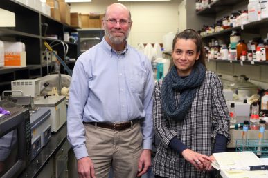
Researchers led by Robert Paul Malchow, associate professor of biological sciences at the University of Illinois at Chicago, have discovered that glial cells may modulate the release of neurotransmitters — chemicals that relay signals between nerve cells — by increasing the acidity of the extracellular environment. Their findings are reported in the online journal PLOS ONE.
Neurons communicate with one another both electrically – through action potentials that result in changes in cell membrane permeability — and chemically through the release of neurotransmitters, such as serotonin and dopamine. Adenosine triphosphate, or ATP, is best known for its role in metabolism where it helps cells use energy, but is also a common neurotransmitter. Previous research has suggested that ATP might play a role in signaling between glial cells and nerve cells.
When the researchers applied ATP to retinal glial cells, they saw an immediate and massive release of acid from the cells. “We decided to further investigate ATP and glial cells because of this really strong response that raised acid levels in the environment right next to the glial cells more than 1000 percent,” said Malchow.
In living organisms, ATP is released by neurons into the space between nerve cells called the synapse, when a nerve cell becomes excited. In other words, when the nerve cell is actively relaying a message to its neighbors.
The researchers determined that a rise in ATP outside the nerve cells causes adjacent glial cells to release hydrogen ions, which raise the acidity of the immediate extracellular environment. The hydrogen ions, in turn, bind to calcium channels in the nerve cell membranes, closing these channels off. The calcium channels, when open, allow for the release of neurotransmitters.
“Increases in acidity in the extracellular environment produced by the glial cells forms a feedback loop which prevents the release of too much neurotransmitter,” said Malchow.
The researchers used ultrasensitive pH sensors developed at the Marine Biological Laboratory to measure the changes in pH levels around isolated retinal glial cells from a variety of organisms, including humans.
The researchers hypothesize that acid released by glial cells decreases the release of neurotransmitters from nerve cells. This is because the acid binds to and inhibits neuronal calcium channels, which control the release of neurotransmitters.
“We believe that this ATP-mediated release of protons by glial cells acts as an essential feedback mechanism throughout the nervous system to limit over-excitability of neurons,” Malchow explained.
Boriana Tchernookova from the University of Illinois at Chicago is the lead author of the paper. Chad Heer, Marin Young, David Swygart, Ryan Kaufman, Michael Gongwer, Lexi Shepherd, Hannah Caringal and Matthew Kreitzer of Indiana Wesleyan University, and Jason Jacoby of the University of Illinois at Chicago, are co-authors on the paper.
This work was supported by National Science Foundation Grants 0924372, 0924383, 1359230, 1557820, and 1557725, a Grants in Aid Award from Sigma Xi, the Scientific Research Society, a Laura and Arthur Colwin Summer Fellowship from the Marine Biological Laboratory, a LAS Award for Faculty in the Natural Sciences from the University of Illinois at Chicago, and a gift from the estate of Mr. John C. Hagensick.

